REF: MEN-2026-0045
Imaging
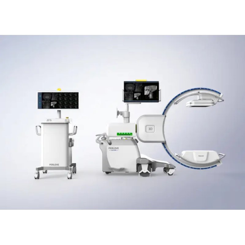
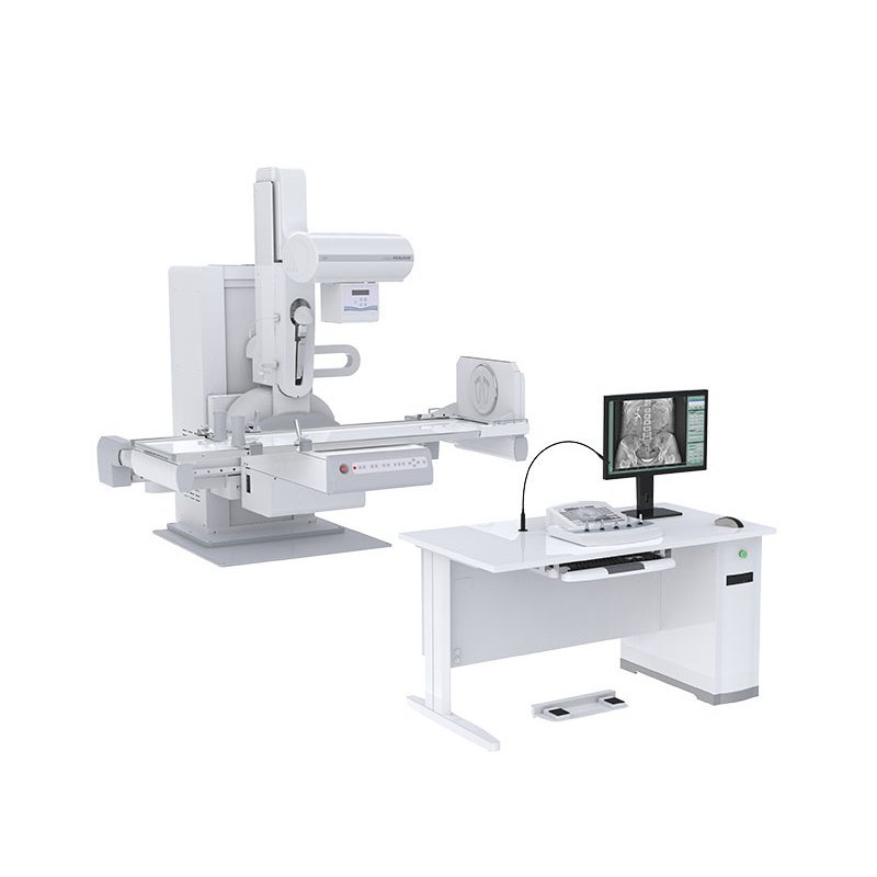
Fluoroscopy Machine
Sold & Shipped By
Official Store
Price on Request
Product Code MEN-2026-0045
Condition New
Availability In Stock
Warranty
Sub-Category Scanning Device
Real-time Imaging: Provides immediate visual feedback during procedures, allowing for quick decision-making and intervention.
High-Quality Images: Utilizes advanced imaging techniques to produce clear and detailed images, essential for accurate diagnosis and treatment.
Low-Dose Imaging: Reduces radiation exposure to patients and healthcare professionals, ensuring safer imaging practices.
Versatile Applications: Supports a wide range of medical procedures, from gastrointestinal studies to orthopedic assessments and angiography.
Mobile and Portable Options: Designed for flexibility in various settings, including operating rooms, emergency rooms, and patient bedside examinations.
Advanced Safety Features: Incorporates technology to manage and reduce radiation exposure, protecting both patients and healthcare providers.
High-Quality Images: Utilizes advanced imaging techniques to produce clear and detailed images, essential for accurate diagnosis and treatment.
Low-Dose Imaging: Reduces radiation exposure to patients and healthcare professionals, ensuring safer imaging practices.
Versatile Applications: Supports a wide range of medical procedures, from gastrointestinal studies to orthopedic assessments and angiography.
Mobile and Portable Options: Designed for flexibility in various settings, including operating rooms, emergency rooms, and patient bedside examinations.
Advanced Safety Features: Incorporates technology to manage and reduce radiation exposure, protecting both patients and healthcare providers.
Please contact our technical team for full specifications.
Preparation and Positioning: The patient is placed on the examination table. The C-arm is moved into position, often requiring the image intensifier to be as close to the patient as possible to reduce magnification, reduce radiation dose, and improve image resolution.
Orbital Rotation: The C-arm rotates around the patient, adjusted by releasing locks on the handle.
Horizontal/Vertical Travel: The boom allows the unit to move forward, backward, or up and down.
Wig-Wag: A 5-degree, side-to-side, or "pan" movement allows for slightly angled imaging, with a larger 10-degree,, or "banana" motion, used for following contrast flow.
Intermittent Imaging: Use short, intermittent bursts of radiation rather than continuous, live imaging to reduce exposure.
Collimation: Narrow the X-ray beam to the specific area of interest to minimize radiation to surrounding tissue.
Last Image Hold (LIH): Use this feature to freeze the last frame for viewing, avoiding unnecessary repeat radiation.
Controls: The operator typically uses a footswitch to activate the X-ray beam, with buttons for printing or saving images.
Radiation Protection: Always wear lead aprons and thyroid shields.
Audible Alarm: The system will sound an alarm every 5 minutes to track beam-on time.
Positioning: Keep the X-ray tube under the table and the image intensifier above the patient to minimize operator dose.
Orbital Rotation: The C-arm rotates around the patient, adjusted by releasing locks on the handle.
Horizontal/Vertical Travel: The boom allows the unit to move forward, backward, or up and down.
Wig-Wag: A 5-degree, side-to-side, or "pan" movement allows for slightly angled imaging, with a larger 10-degree,, or "banana" motion, used for following contrast flow.
Intermittent Imaging: Use short, intermittent bursts of radiation rather than continuous, live imaging to reduce exposure.
Collimation: Narrow the X-ray beam to the specific area of interest to minimize radiation to surrounding tissue.
Last Image Hold (LIH): Use this feature to freeze the last frame for viewing, avoiding unnecessary repeat radiation.
Controls: The operator typically uses a footswitch to activate the X-ray beam, with buttons for printing or saving images.
Radiation Protection: Always wear lead aprons and thyroid shields.
Audible Alarm: The system will sound an alarm every 5 minutes to track beam-on time.
Positioning: Keep the X-ray tube under the table and the image intensifier above the patient to minimize operator dose.