REF: MEN-2026-0042
Imaging
C-arm Machine
Sold & Shipped By
Official Store
Price on Request
Product Code MEN-2026-0042
Condition New
Availability In Stock
Warranty
Sub-Category Scanning Device
Real-Time Imaging & High Resolution: C-arms provide immediate, live, and high-definition X-ray images, allowing surgeons to visualize anatomical structures during procedures, ensuring precision and reduced errors.
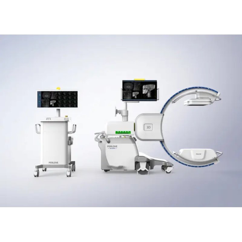
Flexible, Mobile Design: The C-shaped arm connects the X-ray source and detector, enabling 360-degree rotation and flexible positioning (vertical, horizontal, orbital) around the patient to capture multiple angles without moving the patient.
Digital Imaging (Flat Panel Detectors): Modern C-arms often feature digital flat-panel detectors (FPD), which offer superior image quality, wider dynamic range, and lower radiation doses compared to traditional, older intensifiers.
Advanced Imaging Modes: Systems include pulsed fluoroscopy, last-image-hold (LIH), and digital subtraction angiography (DSA) to minimize radiation exposure while optimizing image clarity for specific, intricate tasks.
User-Friendly Interface & Memory: Equipped with intuitive touchscreens and workstations, allowing quick adjustments of settings and digital,,, storage of images for instant review or network sharing.
Versatile Applications: Designed for various medical fields, including orthopedics (fracture fixation), pain management, urology, and cardiac procedures.
Flexible, Mobile Design: The C-shaped arm connects the X-ray source and detector, enabling 360-degree rotation and flexible positioning (vertical, horizontal, orbital) around the patient to capture multiple angles without moving the patient.
Digital Imaging (Flat Panel Detectors): Modern C-arms often feature digital flat-panel detectors (FPD), which offer superior image quality, wider dynamic range, and lower radiation doses compared to traditional, older intensifiers.
Advanced Imaging Modes: Systems include pulsed fluoroscopy, last-image-hold (LIH), and digital subtraction angiography (DSA) to minimize radiation exposure while optimizing image clarity for specific, intricate tasks.
User-Friendly Interface & Memory: Equipped with intuitive touchscreens and workstations, allowing quick adjustments of settings and digital,,, storage of images for instant review or network sharing.
Versatile Applications: Designed for various medical fields, including orthopedics (fracture fixation), pain management, urology, and cardiac procedures.
Please contact our technical team for full specifications.
Setup & Power On: Plug in the machine, turn on the system using the power button (often green), and wait for it to boot up.
Patient Input: Enter patient details on the touch screen or workstation, or select a scheduled case.
Positioning: Unlock the C-arm brakes to move it into position. Place the X-ray tube below the patient and the image intensifier above (or as required) to reduce scatter radiation exposure to staff.
Imaging: Use the foot pedal to take images. A short press usually saves a still image, while holding it enables live, continuous fluoroscopy (sometimes called "cine").
Adjustments: Use the console to adjust image quality (contrast/brightness), invert images, zoom, or rotate them to get the clearest view.
Storage & Shutdown: Save images to the PACS system. Once finished, ensure the system is properly shut down to protect the software.
Key Tips:
Safety First: Always wear lead aprons, thyroid shields, and utilize dosimeters. Keep the image receptor as close to the patient as possible to reduce magnification and radiation scatter.
Controls: The C-arm can be moved in six ways: longitudinal travel, lateral travel, vertical movement, orbital rotation, horizontal scan (wig-wag), and swivel.
Sterile Field: Use sterile covers for the C-arm when working in the surgical field.
Patient Input: Enter patient details on the touch screen or workstation, or select a scheduled case.
Positioning: Unlock the C-arm brakes to move it into position. Place the X-ray tube below the patient and the image intensifier above (or as required) to reduce scatter radiation exposure to staff.
Imaging: Use the foot pedal to take images. A short press usually saves a still image, while holding it enables live, continuous fluoroscopy (sometimes called "cine").
Adjustments: Use the console to adjust image quality (contrast/brightness), invert images, zoom, or rotate them to get the clearest view.
Storage & Shutdown: Save images to the PACS system. Once finished, ensure the system is properly shut down to protect the software.
Key Tips:
Safety First: Always wear lead aprons, thyroid shields, and utilize dosimeters. Keep the image receptor as close to the patient as possible to reduce magnification and radiation scatter.
Controls: The C-arm can be moved in six ways: longitudinal travel, lateral travel, vertical movement, orbital rotation, horizontal scan (wig-wag), and swivel.
Sterile Field: Use sterile covers for the C-arm when working in the surgical field.