Anke Anatom 64 Clarity / Anatom 64 Precision Lower dose and lower consumption to get high quality images World Leading Precision Technology Platform
Following the breakthroughs in the medical system development, Anke proudly introduces the creatively designed ANATOM 64, as a tool of precision medicine in diagnosis imaging. Via the breakthrough designs in precise hardware, software and imaging technologies, ANATOM 64 can provide precise diagnosis information and early detection for small lesions.
Precise hardware, Precise technology, Precise imaging
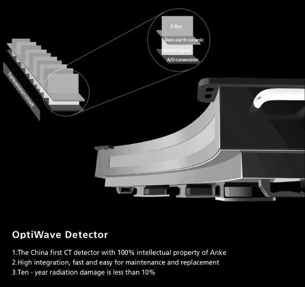
- OptiWave detector
- High precision gantry
control - Dual-mode gantry tilt
- 3D Admir iterative technology
- Dual-energy head imaging
- 1024 x1024 matrix imaging
technology - High-definition imaging
of targeted organs - Low dose platform
- 3D enhanced VR
Precision Technology Platform
ANATOM precision technology platform is equipped with advanced imaging technologies, and adopts 3D OptiWave detector,
Ahead dual-energy imaging: Ahead creatively uses 140kV and 80kV dual energy switching scan mode for brain imaging. By careful analyzing the high and low energy characteristics, images can show more valuable information about the brain tissues
Admir iterative reconstruction technology: Admir applies mathematical and physics models to accurately construct and describe the signal’s quantum characteristics. Iterative operations are performed in the three domains of raw data, projection and image, to greatly reduce the image noise and achieve optimal image quality with low dose.
AccuTilt dual-mode tilt gantry technology: The system provides digital and mechanical tilt to accommodate different user habits and clinical needs. Real-time collision preventing system is available for the patients’ safety, to provide powerful support for accurate diagnosis.
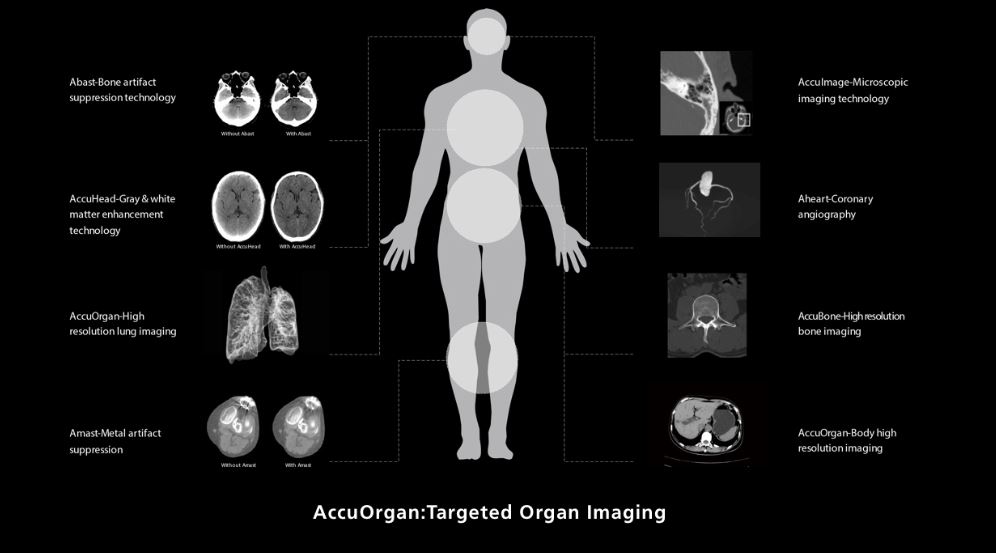
AccuOrgan-Targeted organ imaging: To achieve high precision imaging of each part of human body at low dose and low energy consumption.
Abast-Bone artifact suppression technology: Abast eliminates the X-ray beam hardening effects to the cerebellum, brain stem and other parts of the brain and clearly shows the structure and lesions of the brain stem and cerebellum.
AccuHead-Gray & white matter enhancement technology: AccuHead technology is specifically designed for brain scans to improve the contrast between gray matter and white matter without sacrificing image quality
AccuOrgan-High resolution lung imaging: High resolution images of the lung can be obtained at only 30% ~ 40% of conventional radiation dose
Amast-Metal artifact suppression: Dual-domain iteration is adopted to effectively remove metal artifacts and restore the soft tissue around the metal
AccuImage-Microscopic imaging technology: 1024×1024 matrix to display more details of the pathological changes and provide a reliable information for early detection, early diagnosis and early treatment of the diseases
Aheart-Coronary angiography: Innovative ECG gated trigger and real-time dynamic tube current modulation are applied to achieve low dose HD coronary angiography, which can clearly reveal the fine structure of coronary arteries
AccuBone-High resolution bone imaging: Enhanced bone edge contrast can provide accurate anatomic relationships and show early destruction and cyst of subchondral bone like lesions and articular cartilage calcifications
AccuOrgan-Body high: Combined with the AccuImage microscopic imaging technology, AccuOrgan technology can significantly increase the display of fine structure and morphology of the abdomen and provide more accurate images for the early diagnosis of small lesion
AccuDose-Comprehensive low dose imaging: Pediatric Scan Protocol, Individual Dose Monitoring, AccuShape Filter, Efficient Detector, Adose Dose Modulation, Ahead-Head Dual-energy Imaging, Iterative Reconstruction, Amast, Contrast Agent Tracking
Technology.
AccuScan-Enjoy ease: Convenient and efficient operation process greatly improve work efficiency to achieve high volume of patients
- System: CT scanner
- Application: for whole-body tomography
- Number of slices: 64-slice
-
- easy to use.
- practicality.
- versatility.
- high definition of obtained images.
- high-definition 3D images of the area under study.
- the procedure is pain-free.
- fast data processing.
- possibility to store the image in the computer memory.
- the diagnostics does not take a lot of time.
- low radiation dose.




Reviews
There are no reviews yet.