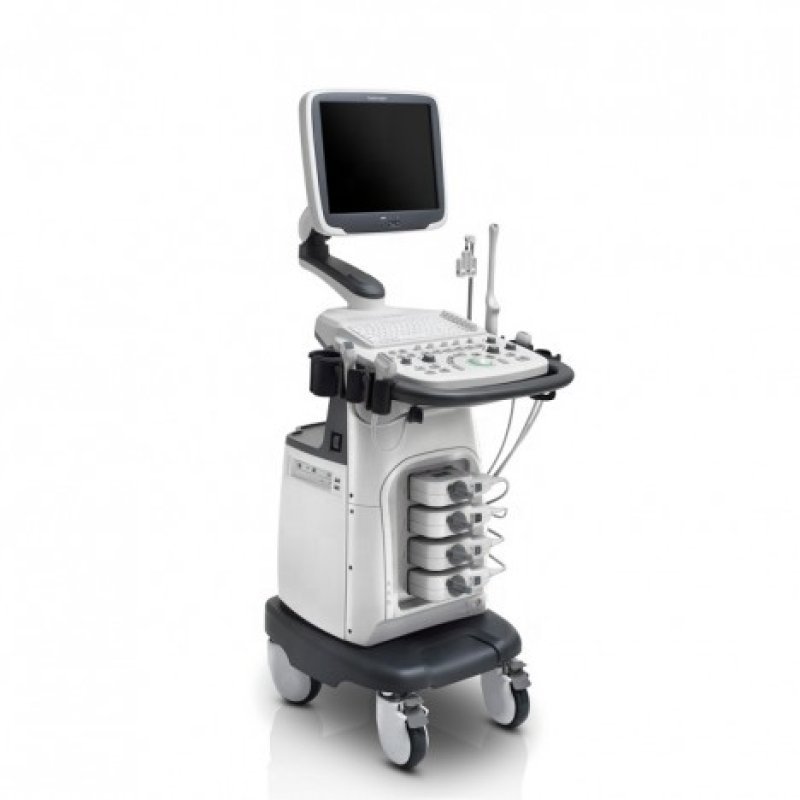
REF: MEN-2026-0047
Radiology
3D/4D Ultrasound
Sold & Shipped By
Official Store
Price on Request
Product Code MEN-2026-0047
Condition New
Availability In Stock
Warranty
Sub-Category scanning machine
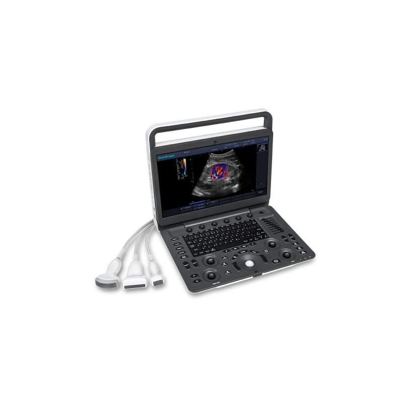
Real-Time Motion Imaging: Unlike 3D, which produces a still image, 4D ultrasound displays movement as it happens, allowing parents to see their baby yawning, stretching, blinking, or smiling.
Volumetric Data Acquisition: The technology uses advanced, high-speed computing to process multiple 2D images, creating a detailed 3D "volume" of the fetus.
Improved Structural Detail: 4D imaging provides clearer views of surface features, such as the face, hands, feet, and umbilical cord.
Fetal Behavior Assessment: It allows doctors to monitor movement patterns to assess neurological development.
Diagnostic Support: While not always necessary, 4D scans can assist in identifying anomalies like cleft lips or skeletal abnormalities.
Best Time for Imaging: The optimal time for a 4D scan is generally between 26 and 30 weeks (or up to 34 weeks) of pregnancy, when the fetus has developed enough facial fat, and there is sufficient amniotic fluid for high-quality images.
Volumetric Data Acquisition: The technology uses advanced, high-speed computing to process multiple 2D images, creating a detailed 3D "volume" of the fetus.
Improved Structural Detail: 4D imaging provides clearer views of surface features, such as the face, hands, feet, and umbilical cord.
Fetal Behavior Assessment: It allows doctors to monitor movement patterns to assess neurological development.
Diagnostic Support: While not always necessary, 4D scans can assist in identifying anomalies like cleft lips or skeletal abnormalities.
Best Time for Imaging: The optimal time for a 4D scan is generally between 26 and 30 weeks (or up to 34 weeks) of pregnancy, when the fetus has developed enough facial fat, and there is sufficient amniotic fluid for high-quality images.
Please contact our technical team for full specifications.
Preparation: Ensure there is adequate amniotic fluid around the fetus. A light snack or fruit juice 15-30 minutes prior can help increase fetal movement.
Initial Setup (2D): Obtain a clear 2D sagittal view (profile) of the fetus, as a good 4D image depends on a solid 2D foundation.
Activate 4D Mode: Press the designated "4D" or "3D/4D" button on the ultrasound machine console.
Adjust Region of Interest (ROI): A yellow box will appear. Use the trackball and "set" key to adjust the size and position of this box to surround only the area of interest (e.g., the face). Making the box smaller improves the frame rate.
Positioning and Rendering: Align the green line, often at the top of the box, parallel to the fetal face to optimize the rendering.
Optimize Image: Adjust the "gain" or "surface" settings to reduce noise and enhance facial features.
Record: Use the "live" or "record" button to capture the real-time movement.
Initial Setup (2D): Obtain a clear 2D sagittal view (profile) of the fetus, as a good 4D image depends on a solid 2D foundation.
Activate 4D Mode: Press the designated "4D" or "3D/4D" button on the ultrasound machine console.
Adjust Region of Interest (ROI): A yellow box will appear. Use the trackball and "set" key to adjust the size and position of this box to surround only the area of interest (e.g., the face). Making the box smaller improves the frame rate.
Positioning and Rendering: Align the green line, often at the top of the box, parallel to the fetal face to optimize the rendering.
Optimize Image: Adjust the "gain" or "surface" settings to reduce noise and enhance facial features.
Record: Use the "live" or "record" button to capture the real-time movement.