REF: MEN-2026-0073
Radiology
Mobile X-ray Machine
Sold & Shipped By
Official Store
₦8,000,000.00
Product Code MEN-2026-0073
Condition New
Availability In Stock
Warranty 1 year
Sub-Category X-ray
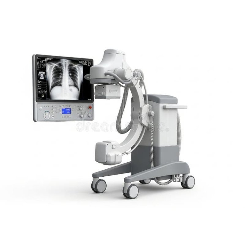
Imaging Capabilities & Power: High-frequency, 5 kW generator (100mA, 40-120kV or 50-90kVp) provides consistent, high-quality images for various diagnostic needs.
Portability & Design: Compact, lightweight, and easy to maneuver, making them ideal for in-hospital, ICU, ER, and, operating room use.
Control Interface: Usually features a digital LCD screen, 50-70+ pre-set, programmable, APR (Anatomical Programmed Radiography) programs, and, simple, soft-touch keypads.
Safety & Protection: Features include analog, MAs digital closed-loop control, automatic error protection, and, beam-limiting devices.
Imaging Software: Often includes, advanced, image processing, software with DICOM 3.0 compatibility for seamless networking, and, high-quality, digital, imaging, capability.
Exposure Control: Remote wireless, exposure, and, hand-switches with extendable, cables, ensure, operator safety.
Power Supply: Generally operates, on single-phase 220V±10% AC, often with, power, factor correction (APFC), integrated, for improved,, energy, efficiency, and, stability.
High Performance: Some models feature advanced digital, flat-panel, detectors, allowing for instant, image, visualization and high, resolution, radiography,.
Portability & Design: Compact, lightweight, and easy to maneuver, making them ideal for in-hospital, ICU, ER, and, operating room use.
Control Interface: Usually features a digital LCD screen, 50-70+ pre-set, programmable, APR (Anatomical Programmed Radiography) programs, and, simple, soft-touch keypads.
Safety & Protection: Features include analog, MAs digital closed-loop control, automatic error protection, and, beam-limiting devices.
Imaging Software: Often includes, advanced, image processing, software with DICOM 3.0 compatibility for seamless networking, and, high-quality, digital, imaging, capability.
Exposure Control: Remote wireless, exposure, and, hand-switches with extendable, cables, ensure, operator safety.
Power Supply: Generally operates, on single-phase 220V±10% AC, often with, power, factor correction (APFC), integrated, for improved,, energy, efficiency, and, stability.
High Performance: Some models feature advanced digital, flat-panel, detectors, allowing for instant, image, visualization and high, resolution, radiography,.
Power Output: Typically 5 kW to 6.4 kW.
High Frequency: Generally ≥40 kHz to 100 kHz, ensuring high-quality X-rays.
mA Range: 10mA to 100mA (often in steps: 16, 32, 63, 100mA).
kVp Range: 40kV to 100kV or 125kV (in 1 kV steps).
mAs Range: 0.1 mAs to 200 mAs or 320 mAs.
Exposure Time: 0.002s to 4s or 0.01s to 6.3s.
Anode Type: Usually stationary anode.
Focal Spot: 0.6mm / 1.8mm (double focus) or 1.5mm.
Anode Heat Capacity: ~40 kHU to 50 kHU.
Detector Type: Wireless or Wired Flat Panel Detector (FPD).
Detector Size: 14" x 17" (35cm x 43cm) or 17" x 17".
Resolution: Spatial resolution ≥3.4 lp/mm.
Pixel Matrix: ≥2304 x 2800 pixels.
Image Acquisition Time: ~3 seconds.
Weight: Approximately 120 kg to 160 kg for the unit, with tube heads weighing roughly 18-35 kg.
Collimator: LED light-based with manual control.
Movements: Counterbalanced, vertical, in-and-out, and angular tube rotation (often ±90° to ±180°).
Power Supply: 220V ±10% AC, 50/60 Hz.
Protection: Overload protection, self-diagnosis, and error alarms.
Compliance: AERB, CE, and ISO 13485 certified.
High Frequency: Generally ≥40 kHz to 100 kHz, ensuring high-quality X-rays.
mA Range: 10mA to 100mA (often in steps: 16, 32, 63, 100mA).
kVp Range: 40kV to 100kV or 125kV (in 1 kV steps).
mAs Range: 0.1 mAs to 200 mAs or 320 mAs.
Exposure Time: 0.002s to 4s or 0.01s to 6.3s.
Anode Type: Usually stationary anode.
Focal Spot: 0.6mm / 1.8mm (double focus) or 1.5mm.
Anode Heat Capacity: ~40 kHU to 50 kHU.
Detector Type: Wireless or Wired Flat Panel Detector (FPD).
Detector Size: 14" x 17" (35cm x 43cm) or 17" x 17".
Resolution: Spatial resolution ≥3.4 lp/mm.
Pixel Matrix: ≥2304 x 2800 pixels.
Image Acquisition Time: ~3 seconds.
Weight: Approximately 120 kg to 160 kg for the unit, with tube heads weighing roughly 18-35 kg.
Collimator: LED light-based with manual control.
Movements: Counterbalanced, vertical, in-and-out, and angular tube rotation (often ±90° to ±180°).
Power Supply: 220V ±10% AC, 50/60 Hz.
Protection: Overload protection, self-diagnosis, and error alarms.
Compliance: AERB, CE, and ISO 13485 certified.
Bedside Imaging in ICUs/Wards: Used in Intensive Care Units and hospital wards for critically ill or immobile patients, reducing the risks associated with transporting them to a radiology department.
Emergency & Trauma Rooms: Provides immediate diagnostic images for trauma cases, such as fractures or dislocations, aiding in rapid assessment and treatment.
Operating Rooms (OR): Utilized for intraoperative imaging, such as verifying the correct placement of surgical hardware or markers.
Orthopedic Evaluations: Used in orthopedic clinics for visualizing bone and joint structures for diagnosis, treatment planning, and post-operative monitoring.
Field & Disaster Response: Ideal for use in temporary, rural, or remote clinics, including war zones or disaster areas, due to its compact and mobile nature.
Veterinary Medicine: Employed for both small and large animal diagnostics in clinics or on-site, such as equine imaging. Pediatric Care: Allows for imaging in familiar, less stressful environments, with adjustable doses to minimize radiation exposure for children.
Emergency & Trauma Rooms: Provides immediate diagnostic images for trauma cases, such as fractures or dislocations, aiding in rapid assessment and treatment.
Operating Rooms (OR): Utilized for intraoperative imaging, such as verifying the correct placement of surgical hardware or markers.
Orthopedic Evaluations: Used in orthopedic clinics for visualizing bone and joint structures for diagnosis, treatment planning, and post-operative monitoring.
Field & Disaster Response: Ideal for use in temporary, rural, or remote clinics, including war zones or disaster areas, due to its compact and mobile nature.
Veterinary Medicine: Employed for both small and large animal diagnostics in clinics or on-site, such as equine imaging. Pediatric Care: Allows for imaging in familiar, less stressful environments, with adjustable doses to minimize radiation exposure for children.