REF: MEN-2026-0284
Gastroenterology
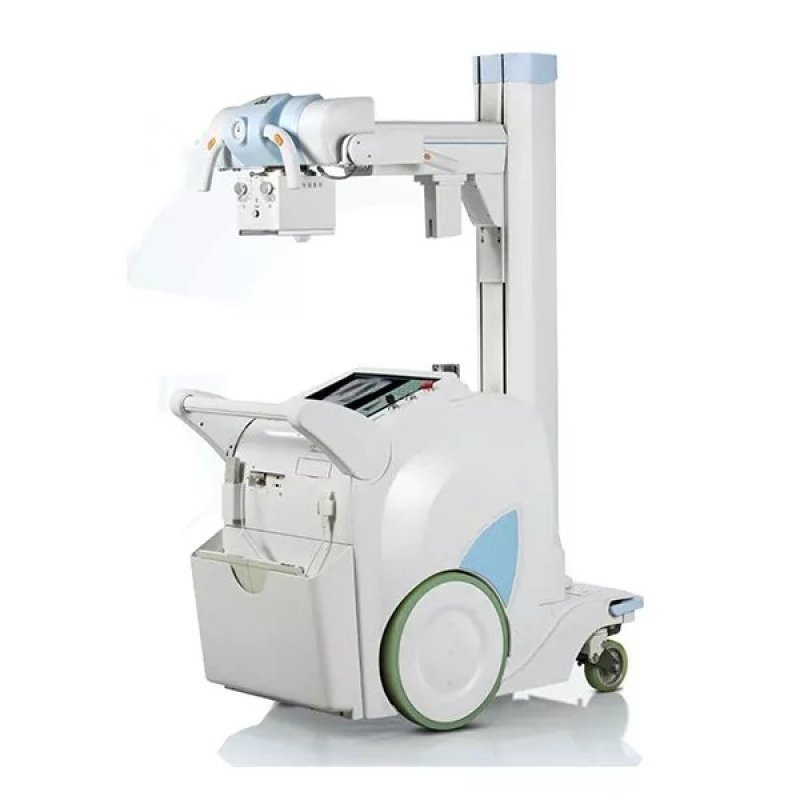
Mobile Digital X-Ray System Plx- 6200
Sold & Shipped By
Official Store
Price on Request
Product Code MEN-2026-0284
Condition New
Availability On request
Warranty
Sub-Category Imaging Device
.1
standard output power: 32 kW; max output power: 40 kW
1.2
mA range: 10-400 mA
1.3
Tube voltage range: 40-150 kV
1.4
Mass range: 0.1-500m As
1.5
Load time range: 0.001s~6.3s
1.6
Input voltage: 336VDC±10%
1.7
High-voltage transformer structure: The power frequency
standard output power: 32 kW; max output power: 40 kW
1.2
mA range: 10-400 mA
1.3
Tube voltage range: 40-150 kV
1.4
Mass range: 0.1-500m As
1.5
Load time range: 0.001s~6.3s
1.6
Input voltage: 336VDC±10%
1.7
High-voltage transformer structure: The power frequency
X-ray high-voltage generator
1.1 standard output power: 32 kW max output power: 40 kW
1.2 mA range: 10-400 mA
1.3 Tube voltage range: 40-150 kV
1.4 mAs range: 0.1-500m As
1.5 Load time range: 0.001s~6.3s
1.6 Input voltage: 336VDC±10%
1.7 High-voltage transformer structure: The power frequency
2. X-ray tube
2.1 Focus range: 0.6/1.2 mm
2.2 Anode heat capacity: 300k Hu
2.3 Pipe sleeve heat capacity: 1250 kHu/900 kJ
2.4 The maximum voltage: 150 kV
2.4 The anode input power: Big focus: 50 kW, Small focus: 22 kW
2.5 Anode angle 12°
2.6 The inherent filtration: 1.3 mm Al/75 kV
3. Collimator
3.1 The inherent filtration: 1.2 mm Al 70 kV;
3.2 Collimator is equipped with LED lights (Bright 30s are automatically extinguished).
3.3 Collimator is built into measuring ruler
4. The tube bracket structure
4.1 X-ray tube support way: cross arm
4.2 Tube focus distance to the ground: 570 mm~1800 mm, ±5 %
4.3 Telescopic Arm Length Range: 710 mm~1240 mm ㎜
4.4 Column Rotation Range: ±270°
4.5 Tube Tilting Angle: ±180°
4.6 Tube Vertical Rotation Angle: -30°~+90°
4.7 Collimator axial rotation: ±90°
5. Detector
5.1 Detector: Pax Scan 4336W
5.2 WIFI connection, amorphous silicon, whole panel technology
5.3 Detector valid exposure size: 427×344 mm
5.4 Pixel size: 150 µm
5.5 The effective camera size of the detector: 14×17 inches
5.6 Gathering matrix: 2304×2800 pixels;
5.7 Spatial resolution: 3.4 lp/mm
5.8 Pixel gray level: ≥14 bit
5.9 Detector cooling mode: natural cooling
5.10 Imaging time: ≤ 5 s
6. Digital image workstation MOC (V02.01.00)
6.1 CPU frequency: CORE i5
6.2 Memory capacity: 4GB
6.3 Hard drive capacity: 500 GB
6.4 Display size: 19-inch LCD touch screen
6.5 Display screen maximum resolution ≥ 1280×1024;
6.6 64-bit Windows operation system
7. Image acquisition software
7.1 Patient registration: acquisition, find the patient data, to prepare for the photography
7.2 Radiographic examination: setting system parameters, expose photography, and get the image
7.3 Images display, image analysis, processing and output
7.4 Data management: managing patient data, leading in, leading out , batch file, etc. in accordance with DICOM 3.0
7.5 Configure calibration: the system components such as flat-panel detector, such as the tube, for automatic configuration and calibration
7.6 System diagnosis: monitoring system status, diagnosis of the system fault
8. Battery
8.1 Power supply way
8.1.1 storage battery supply
8.1.2 Battery capacity: Maximum 240 exposures, each time mAs, chest image dosage (exposure conditions: 120 kv, 100 mA, ms)
8.2 There is no wall charge when exposed
8.3 Can support hand switch and remote controls
9. The machine parameters
9.1 Mobile mode: infinitely variable speed
9.2 Working mode: intermittent loading, continuous operation
9.3 Appearance size
9.3.1 Whole machine width: 670 mm
9.3.2 Whole machine length: 1235 mm
9.3.3 Whole machine height: 1825 mm
9.4 Whole weight: 518 kg
9.5 Drive mode: dual motor drive, respectively control left and right driving wheel
9.6 Collision prevention: obstacle contact sensor
10. External interface
10.1 Dual display interface (VGA/DVI)
10.2 Wireless network data transmission (WIFI)
10.3 Cable network interface
10.4 USB (4 units)
10.5 Choose a chest rack
10.6 The selection of moving flatbed
11. After-sales service
11.1 The machine warranty 1 year
11.2 1 set of complete operation and maintenance manual
11.3 Provide site application and service training
11.4 Free service hotline
12. copyright certificate
12.1 DR service control application software
12.2 mobile Dr embedded system software
12.3 mobile Dr generator control software
1.1 standard output power: 32 kW max output power: 40 kW
1.2 mA range: 10-400 mA
1.3 Tube voltage range: 40-150 kV
1.4 mAs range: 0.1-500m As
1.5 Load time range: 0.001s~6.3s
1.6 Input voltage: 336VDC±10%
1.7 High-voltage transformer structure: The power frequency
2. X-ray tube
2.1 Focus range: 0.6/1.2 mm
2.2 Anode heat capacity: 300k Hu
2.3 Pipe sleeve heat capacity: 1250 kHu/900 kJ
2.4 The maximum voltage: 150 kV
2.4 The anode input power: Big focus: 50 kW, Small focus: 22 kW
2.5 Anode angle 12°
2.6 The inherent filtration: 1.3 mm Al/75 kV
3. Collimator
3.1 The inherent filtration: 1.2 mm Al 70 kV;
3.2 Collimator is equipped with LED lights (Bright 30s are automatically extinguished).
3.3 Collimator is built into measuring ruler
4. The tube bracket structure
4.1 X-ray tube support way: cross arm
4.2 Tube focus distance to the ground: 570 mm~1800 mm, ±5 %
4.3 Telescopic Arm Length Range: 710 mm~1240 mm ㎜
4.4 Column Rotation Range: ±270°
4.5 Tube Tilting Angle: ±180°
4.6 Tube Vertical Rotation Angle: -30°~+90°
4.7 Collimator axial rotation: ±90°
5. Detector
5.1 Detector: Pax Scan 4336W
5.2 WIFI connection, amorphous silicon, whole panel technology
5.3 Detector valid exposure size: 427×344 mm
5.4 Pixel size: 150 µm
5.5 The effective camera size of the detector: 14×17 inches
5.6 Gathering matrix: 2304×2800 pixels;
5.7 Spatial resolution: 3.4 lp/mm
5.8 Pixel gray level: ≥14 bit
5.9 Detector cooling mode: natural cooling
5.10 Imaging time: ≤ 5 s
6. Digital image workstation MOC (V02.01.00)
6.1 CPU frequency: CORE i5
6.2 Memory capacity: 4GB
6.3 Hard drive capacity: 500 GB
6.4 Display size: 19-inch LCD touch screen
6.5 Display screen maximum resolution ≥ 1280×1024;
6.6 64-bit Windows operation system
7. Image acquisition software
7.1 Patient registration: acquisition, find the patient data, to prepare for the photography
7.2 Radiographic examination: setting system parameters, expose photography, and get the image
7.3 Images display, image analysis, processing and output
7.4 Data management: managing patient data, leading in, leading out , batch file, etc. in accordance with DICOM 3.0
7.5 Configure calibration: the system components such as flat-panel detector, such as the tube, for automatic configuration and calibration
7.6 System diagnosis: monitoring system status, diagnosis of the system fault
8. Battery
8.1 Power supply way
8.1.1 storage battery supply
8.1.2 Battery capacity: Maximum 240 exposures, each time mAs, chest image dosage (exposure conditions: 120 kv, 100 mA, ms)
8.2 There is no wall charge when exposed
8.3 Can support hand switch and remote controls
9. The machine parameters
9.1 Mobile mode: infinitely variable speed
9.2 Working mode: intermittent loading, continuous operation
9.3 Appearance size
9.3.1 Whole machine width: 670 mm
9.3.2 Whole machine length: 1235 mm
9.3.3 Whole machine height: 1825 mm
9.4 Whole weight: 518 kg
9.5 Drive mode: dual motor drive, respectively control left and right driving wheel
9.6 Collision prevention: obstacle contact sensor
10. External interface
10.1 Dual display interface (VGA/DVI)
10.2 Wireless network data transmission (WIFI)
10.3 Cable network interface
10.4 USB (4 units)
10.5 Choose a chest rack
10.6 The selection of moving flatbed
11. After-sales service
11.1 The machine warranty 1 year
11.2 1 set of complete operation and maintenance manual
11.3 Provide site application and service training
11.4 Free service hotline
12. copyright certificate
12.1 DR service control application software
12.2 mobile Dr embedded system software
12.3 mobile Dr generator control software
Operation manuals are available upon request during delivery.